Raising public interest in the microscopic world
Feedbacks from my experience at the 2018 European researcher's night
I took part in the 2018 edition of the European Researcher’s Night, an outreach event happening every year all over Europe at the end of September, and funded by the Marie Curie Actions. I joined some of my colleagues from my research institute, the Centre for Organismal Studies (COS), to animate an activity around microscopy and the observation of the microscopic world, using microscopes and Foldscopes. This outreach activity was the occasion for me to share my interest in microscopy. 🔬
Outreach is part of my job as a researcher. Funding that supports research projects is mainly, if not entirely, coming from public money, in other words the taxes that each of us pay. Hence, I think it is a duty to communicate about how we, researchers, use this money. I also think that it is a duty to share our knowledge and raise the interest of the public for science. I personally enjoy a lot outreach, and see it as a chance to take a step back on my research subject. I try to get involved in outreach activities at least once a year.
A great outreach experience at the European researcher’s night 2018
I participated in an event organised by Dr. Michael Raissig during the European Researcher’s Night at the botanical garden in Heidelberg. The event was called “Nature magnified – on your smartphone”. There was four “Foldscope stations”, where people could look at microscopy slides using a Foldscope, and two other stations, where people could have a look at living aquatic animals (hydra and the sea anemone aptaisia), that are studied at our research institute, with the microscopes that we use for our research.
I joined this event to share my interest about microscopy and the observation of the microscopic world with the public, as well as to try Foldscopes. I helped preparing the room the day before, and I animated alone one of the Foldscope stations during two hours on the day of the event. I had at my disposal ten Foldscopes and a dozen of microscopy slides with samples of plants and animals prepared by Dr. Michael Raissig, Dr. Sergio Acebrón and some of my colleagues at the Centre for Organismal Studies (COS), as well as one commercial slide. I enjoyed a lot participating to this event. A lot of people of all ages came to try the Foldscopes and look at the different microscopy slides. I was happy to see that, whether one is researcher or not, we all have the same impatience to discover what is on a microscopy slide, and the same satisfaction when we manage to centre the sample and focus on it. I was also amazed by the robustness of the Foldscopes to so much manipulations and by their simplicity of use.
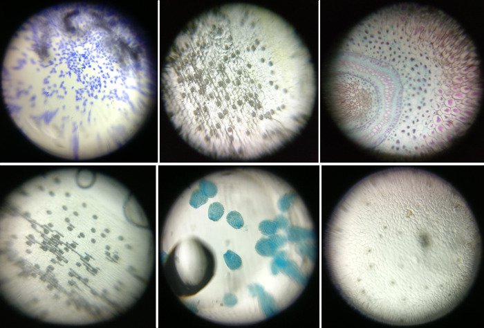
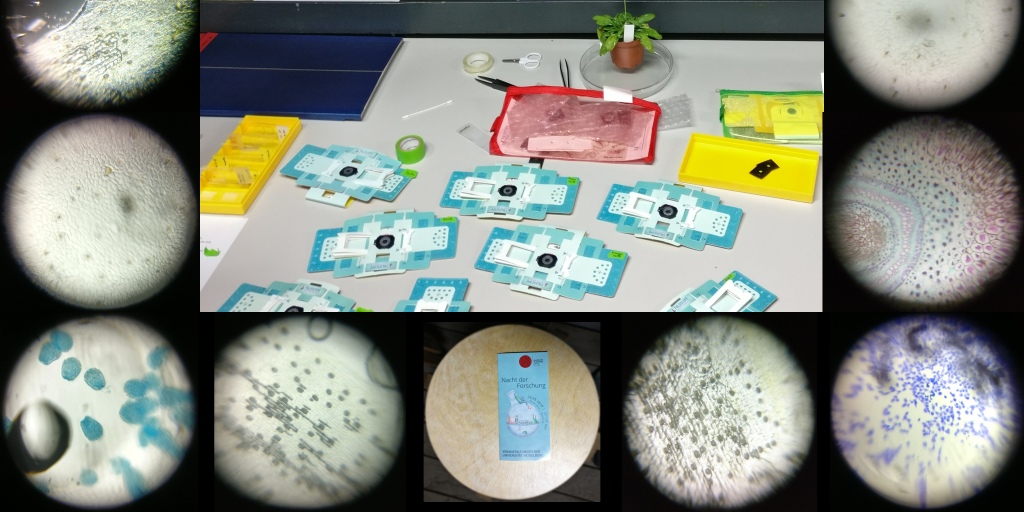
Figure 1: Several microscopy slides viewed with a Foldscope and photographed with a smartphone. From left to right and top to bottom: human cells, plant epidermis, fern rhizome (commercial microscopy slide), plant epidermis, aptaisia larvae (sea anemone), and plant epidermis.
Preparing an outreach activity about microscopy (using Foldscopes)
I would like to document below some elements of the preparation of this event and gather some of my thoughts to help others, as well as me, to re-do it another year. most of the comments below apply to outreach activity with any type of microscopes. However, the main advantages of Foldscopes over other types of microscopes are their low cost and their robustness. It is hence possible to have a large number of them for an outreach event, without the fear of breaking one, and to offer the possibility to all the visitors to observe the microscopic world. Along with the Foldscopes, we also had the chance to have a few “real” microscopes, which offers the possibility to show even more things to the public and notably living animals, like the hydra.
Before the event
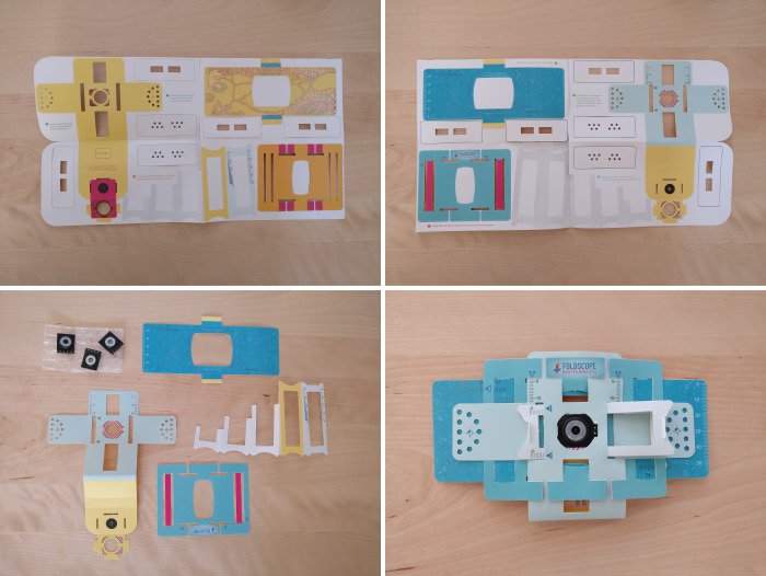
Figure 2: Folding a Foldscope.
- Fold all Foldscopes. Dr. Heike Lindner folded around 40 of them! She said that it took her around 20 minutes for the first ones and only 5 minutes for the last ones. We tried folding one each ourselves during the preparation of the room and it took us 25 to 30 minutes…
- Prepare all the microscopy slides. We had more than 50 homemade microscopy slides in total, with around 8-16 for each kind of samples. These slides were prepared by Dr. Michael Raissig, Dr. Sergio Acebrón and other colleagues at the COS. Two important remarks about the slides:
- All microscopy slides must be perfectly sealed. Inserting them in the Foldscope can be hard the first time as the material composing the Foldscope is new, especially if you use glass slides. I accidentally peeled off the tape of one of the homemade microscopy slides because of this…
- Not all samples are suitable for observation with the Foldscope, nor for this kind of outreach event. As many outreach events were taking place at the same time on the campus of the university where the botanical garden is located, the attention span of the audience was rather short. It is better to choose samples that are neither too small (not easy to see or to find on the slide) or too big (not fitting in the field of view of the Foldscope), nor too homogeneous (uniform colour with few visible details, or no colour). If your own research samples do not match these criteria, do not hesitate to choose other samples. For instance, Dr. Michael Raissig did not show Arabidopsis leaf epidermis as the cells are too small, but rather Amaryllis leaf epidermis. Ideally, it is also better to show samples for which you can also show the whole organism alive or on a picture. For instance, here, the public could observe alive aptaisia at different stages of development and hydra on the nearby microscopy stands, after having seen them with the Foldscopes on slides.
- All microscopy slides must be perfectly sealed. Inserting them in the Foldscope can be hard the first time as the material composing the Foldscope is new, especially if you use glass slides. I accidentally peeled off the tape of one of the homemade microscopy slides because of this…
- Ensure to have at least one commercial slide per station as a plan B in case the other slides are not good enough. Commercial slides are perfectly sealed and usually nicely stained and prepared. I had a slide with a beautiful cross section of a fern rhizome.
One day before the event
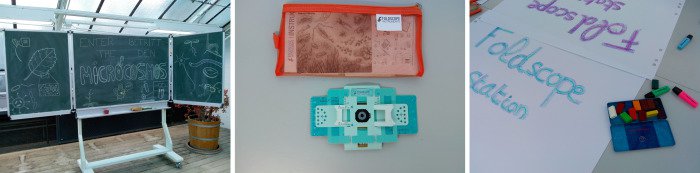
Figure 3: Getting ready for the event.
- Prepare the room. We were located in an old and very nice glasshouse from the botanical garden 🌱 😃 🌱. The day before we were around 10 helpers from the Centre of Organismal Studies (COS). We prepared a blackboard at the entrance with the title of the event, and one table per station with (hand written) A0 posters to indicate the different stations. We also got familiar with the Foldscopes.
On the day of the event
- Finish to install the room. The stations with the standard microscopes were installed only on the day of the event. The living aquatic organisms (hydra and aptaisia), as well as a projector and a computer to project nice microscopy images from the research conducted at our institute were brought just before the event.
- Check all the microscopy slides, if it has not be done yet, and select the best ones. If you have roughly the same number of slides as Foldscopes, you can insert them in the Foldscopes and do the focus before the beginning of the event to spare time. You can also already take a few pictures of each sample with your smartphone. Its help to explain what is on the slides to the public.
- Have fun! 🎉 🔬 🎉
Enjoying the botanic garden of Heidelberg by night @Science_NightHD @cellnetworks @UniHeidelberg #microCOSmos pic.twitter.com/tAUmQWdgSN
— Sergio Perez Acebron (@Acebron) September 28, 2018
#microCOSmos in full swing! @Science_NightHD @UniHeidelberg pic.twitter.com/1GJpWmrKas
— Karin Schumacher (@SchumacherLab) September 28, 2018
It was a great night with microscopes and cocktails in the botanic garden. Good attendance and a lot of fun. Thanks to @MichaelRaissig , Ulrike Engel, all the students, and the workers of the botanic. @Science_NightHD #microCOSmos pic.twitter.com/mFnRfc2UYv
— Sergio Perez Acebron (@Acebron) September 28, 2018
References
About the Foldscope:
https://www.foldscope.com/our-story/
https://journals.plos.org/plosone/article?id=10.1371%2Fjournal.pone.0098781
About this outreach event:
https://en.nacht-der-forschung-heidelberg.de/event/natur-sichtbar-gemacht-auf-deinem-smartphone/
About the European researcher’s night in general:
https://ec.europa.eu/research/mariecurieactions/actions/european-researchers-night_en
Acknowlegdments
I would like to thank Dr. Michael Raissig for taking charge of the organization of the event in coordination with the botanical garden, Dr. Heike Lindner for the preparation of this event, in particular for folding all the Foldscopes and her help for the installation of the room, Dr. Ulrike Engel, from the Nikon imaging centre, for bringing some “real” microscopes and animating one stand on hydra, the members from the lab of Dr. Annika Guse for the animation of the other microscopy stand on aptaisia and the preparation of microscopy slides, and also Dr. Sergio Acebrón and his group, and all the COS members involved in the preparation and animation of the event, as well as the botanical garden of Heidelberg for hosting us. I would also like to thank Dr. Sébastien Rochette (StatnMap; R, models and spatial things) for the pictures of the microscopy slides with a smartphone and the help with inserting the tweets in this article.
Citation:
For attribution, please cite this work as:
Louveaux M. (2018, Oct. 03). "Raising public interest in the microscopic world". Retrieved from https://marionlouveaux.fr/blog/raising-interest-in-the-microscopic-world/.
@misc{Louve2018Raisi,
    author = {Louveaux M},
    title = {Raising public interest in the microscopic world},
    url = {https://marionlouveaux.fr/blog/raising-interest-in-the-microscopic-world/},
    year = {2018}
  }
Share this post
Twitter
Google+
Facebook
LinkedIn
Email